Showing posts with label breast augmentation. Show all posts

Monday, April 29, 2013

Breastfeeding Using Implant Breasts

Breastfeeding Function

Breastfeeding Using Implant BreastsBreasts are basically a milk-producing glands which are used to feed babies. There is a nipple in an aerola area (or called nipple areola complex—NAC), with varying colors from pink to dark brown. Within this gland, breast milk is produced by lactiferous ducts and distributed throughout the breast. For every breast, there are four until eighteen lactiferous ducts ending to the nipple. The comparison of glands to fat within the breasts tissue is 2 to 1—in lactating women—or 1 to 1—in non-lactating women. Thre are more than just glands inside a woman’s breasts; there are collagen, elastin, fat, and ligaments. There are also nerve system in breasts, where the anterior and lateral branches of the fourth, fifth, and sixth nerves are located. Thoraric spinal nerve 4 or T4 in breasts also supplies a specific sensation to the nipple area.


The most important concerns about breastfeeding is in the potential of digestive contamination and toxicity. If the filler of breast implant device is leaked to the breast milk, it will endanger the baby. Substances contained in a breast implant filler is chemically and biologically inert, because they were made of environmentally common substances like salt water (for the saline filler)—although silicone in the filler is unable to be digested. Besides, experts have said that whatever the reasons, there shouldnot be any contraindication for breastfeeding by women with implanted breasts. In the beginning of the use of breast implant (at early 1990s), perhaps there are many non-technical complains from patients and doctors about possible complications from the implant device. Yet, there is no disease casuality related to the device.

Augmented Breasts

Meanwhile, women with implanted breasts are still able to feed babies using their breasts. But, this implant devices may give a kind of difficulties. Mammoplasty surgery, which includes a periareolar incisions and subglandular replacement, is the main cause of this difficulties. Moreover, other difficulties are about the potential damage of lactiferous ducts and the nerves around the nipple area.

If the surgeon cut the ducts and any major nerves within the breast tissue or if the glands are damaged somehow, possible difficulties risk arises. The first is common to surgical procedures that involve periareolar incision implantation because it cuts breast tissue close to the nipple. In the other hand, other implantation incisions like inframmamory fold, trans-axillary breast augmentation, or trans-umbilical breast augmentation, avoid this step. However, if the patient is serious about the possibilities of this breastfeeding difficulties, she can ask the doctor to make effective the incision step so that the damage of the milk ducts and nerves can be reduced. Basically, only implants that are placed under the gland (or called subglandular implants) and implants for large-sized breast that mostly affect the milk glands. Implantation for small size breasts and for submuscle gives less risk of breastfeeding function problems.

Breast Implant Surgical Procedures


Breast implantation, or medically termed as mammoplasty, is a procedure to implant a breast implant device within the breast tissue. There are three purposes of this breast surgery: (1) reconstruction, if the breast tissues were damaged by trauma, disease, or other accidents; (2) revision, to fix up the result of other breast surgeries performed previously; (3) cosmetic purposes, to give the breasts an aesthetically better look.
Breast implant

Five Kinds of Incisions

Breast implant emplacement is performed with five (5) types of surgical incisions:
There are five kinds of surgical incisions in this breast surgery:
  • Inframammary. This is the incision created around the infra-mammary fold (IMF). This incision will give maximum access to most areas within the breast tissues. Because of that, this incision is usually made if the surgeons want to do an emplacement of the silicone-gel breast implants. However, this incision leaves a thicker, more visible scars onto the breast skin.
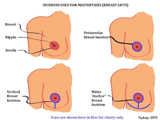
  • Periareolar. This is the incision created in a border line of the areola. This incision will give a better depth-look into the breast tissue right if inframammary incisions need to be adjusted in certain ways. This incision is also necessary to be created if the surgery includes a breast lift (mastopexy). Unlike the inframammary incision, periareolar incision does not enable surgeons to emplace silicone gel implants due to the small size of the incision. Lucky that this incision is less visible than the inframammary incision because it is located around the areola’s border.
  • Transaxillary. This is the incision created in the armpit (axilla). This incision will create a dissection tunnels from where surgeons can emplace the implants. The emplacement can be done both bluntly or with the help of video microcamera, and therefore produces less or even no scars on the breast skin. However, this incision is less acurate in positioning the implant device. That’s why, transaxillary incision in mammoplasty is mostly revised with an inframammary or periareolar incision.
  • Transumbilical. This is the incision created at the navel. This incision is actually less common because it creates a dissection tunnels upwards from the bust. It is also so much trickier to position the breast implant device more accurately, although it creates less or even no scars. Transumbilical incision is not suitable to placing silicone gel implants within the breast tissue because of its inaccuracy nature. Moreover, silicone gel implants cannot be inserted through an incision this small because they are more incompressible.
  • Transabdominal. This is mostly similar to transumbilical incision. This incision creates tunnels from the abdominal incision to the implant tissue. Mostly, an abdominoplasty is undergone simultaneously with this incision.

Breast Implant Emplacement

By definition, breast implant emplacement is a cross-sectional plan of two different implantantions: subglandular breast prosthesis implantation and submuscular breast prosthesis implantations. There are four different procedures can be apply to perform a breast implant emplacement to the implant pocket within the breast tissue.
  • Subglandular. The emplacement is done onto the retromammary area, which is located between the gland and the pectoralis muscle. This emplacement method gives the most good-looking results. However, subglandular position often shows up some ripples and wrinkles, especially in patients with thin pectoral tissue size. This emplacement procedure also gives higher rate of capsular contracture incidence.
  • Subfascial. The emplacement is done under the fascia of the pectoralis muscle. Actually, subfascial emplacement is a variation from subglandular position. This emplacement method is still under debate, because some surgeons said that this emplacement method gives a more vast coverage of implantation and better position sustainability, while other surgeons were against this opinion.
  • Subpectoral. This emplacement is done under the pectoralis muscle. Because this emplacement can only be done after the surgeon wears off the attachments from the inferior muscle, the upper part of the implant is put under the pectoralis muscle while the lower part is in the subglandular area. Although in some patients the possibility of the implants moving from its initial position is a bit higher, subpectoral emplacement cover the maximum area of the implant and allows the lower part of the implant to be expanded.
  • Submuscular. This emplacement is done under the pectoralis muscle, similar to subpectoral emplacement. But, submuscular emplacement does not require the surgeons to wear off the attachments of the inferior muscle. Coverage of this emplacement method can be reached maximally by wearing off the lateral muscle from the chest (the serratus muscle, the pectoralis minor muscle, or the two). In most breast surgery, submuscular emplacement method gives the best result to the breast implants.

Post-surgical recovery

Scars from breast augmentation surgery usually appear 6 weeks after the surgery. It then fades in several months. However, patients can go back to their normal daily life just in a week after surgery as long as they do not do hard physical activities. However, longer recovery time can happen for patients of submuscular placement breast augmentation. These patients can only be back to their daily activities after more than 6 weeks. This is because the incisions of the chest muscles takes longer period to be healed. During this recovery time, it is recommended for the patient to do some light exercises on their arms. These arm exercises are good to let go the pain within the breast tissue. Sometimes, analgesic medication catheters are used to alleviate the pain. Some other advanced techniques of breast implantation enables the patient to recover a lot faster. About 95 percent of women undergoing these techniques can resume their normal lifestyle just in 24 hours with barely any helps of bandages, catheters, and other medical devices.

Wednesday, April 24, 2013

Surgical Procedures of Mastopexy


Surgical Procedures of Mastopexy

Mastopexy for Women

Before performing a mastopexy, surgeons will see if the woman who wants to do this breast-lift surgery comprehends the medical benefits and risks of the procedure. She needs to understand that there are certain body images that can be and cannot be achieved from mastopexy. Some of the indications before running a mastopexy are sagging breasts, post-explantation ptosis, congenital ptosis and pseudoptosis, and acquired or relative ptosis,

For the sagging breast

Mastopexy of the sagging breasts are inquired for both full breast- and modified breast-lift. This technique can only be performed to the surgical incisions to the skin envelope of the breast, not the parenchyma (or the inner substance of the breast).

For a full breast lift, the sagging breasts are lifted under circumvertical and horizontal incision from the Anchor mastopexy. This includes three kinds of incisions: (1) the Anchor ring, an circle-shaped incision on the upper part of the nipple-areola complex; (2) the Anchor shank, a vertical incision from the lower part of the nipple area until the inframammary incision; (3) the Anchor stock, a horizontal incision around the inframammary or around the joint between breast and chest.

In cutting the folds of excess skin from the sagging, inelastic skin-envelope of the breast (and occasionally reducing the nipple-areola complex diameter), the three-incision technique of the Anchor mastopexy allows maximal corrections to the breasts, thereby producing an elevated bust with breasts of natural size, look, and feel. Moreover, each of the three scars to the breast hemisphere produced by the Anchor-pattern mastopexy has a characteristic healing pattern:

To cut the excess skin created from this process (which acquires the sagging skin envelope of the breast, sometimes includes the nipple area diameter), these three kinds of incision permit some maximum corrections of the breasts. Then, it produces an elevated breasts with natural size and appearance. And removing the scars on the breasts due to the mastopexy process requires a skin pigment transition from light to dark skin color—light skin for the breast and dark skin for the areola skin. This is performed for scars around the periareolar area. Meanwhile, for scars on the medial vertical around the nipple areola complex to the inframammary fold or horizontal scars around it, there is no concealing needed because the scars are hidden by the shadow of the breast.

Post-surgically, of the three breast-lift surgery scars, the scar to the inframammary fold exhibits the greatest tendency to hypertrophy, to thickness and large size. Although the coloration of mastopexy scars fades with the full maturation of the tissues, they do remain visible.

In mastopexy for modified breast lift, it takes smaller number of cuts and scars. However, it does not allow the surgeon to make more changes to the skin envelope of the breast. In most occurences, modified breast lift is regarded as a sub-ordinate surgery under a mastopexy for breast augmentation surgery. Sometimes it is also included after lifting and enlarging breast surgery. Some variations of incisions in the modified breast lift are:
  • periareolar lift, with a crescent shaped incision above and at some part of the nipple area, will cut and remove the crescent flesh so that the transposition of the nipple will be a bit higher compared to the breast;
  • circumareolar lift, with a cut on the concentric ring flesh around the nipple area, will prohibit the size of the circle-shaped scar to be maximum;
  • circumvertical lift, with a circumareolar incision around the circle of nipple area and a vertical incision starting from the lower part of the nipple until the inframammary fold.

For the augmented breast

Breast augmentation is sometimes followed by a high potential of breast ptosis. Breast ptosis may be created by stresses—both mechanically and physically—from the implanted breast to the tissue and skin envelope. The overstrecthing thins of the skins may result in the stresses. But, however, based on statistic, breast augmentation and mastopexy come with low medical risks, although the risks may increase of the two are performed altogether and as a combination. The risks can increase the potential of infection of the incision, exposure to the implanted breast, breast and nipple nerves damage, and deformity of nipple and breast implant. The reason of this risk is that breast augmentation and mastopexy, when performed as a combination, may increase the surgical complication level—at least compared to if each is performed separately. Technologies still make several advancement to enable the simultaneous procedure of breast augmentation and mastopexy with a lower risk and medical complication. This advancement, named SAM—stands for simultaneous augmentation mastopexy, includes invaginating and tacking the tissue as the first step. It enables surgeon to previsualize the final result of the surgery before creating any kinds of incision to the breast.

Contraindications

Although most mastopexies are safe, there are certain contraindications appear in some surgical process. These contraindications include aspirin, tobacco smoking, diabetes, and obesity. Surgeon evaluates the fit of a woman who wants to undego a breast lift procedure after explantation, especially for woman with encapsulated breast implants. This facilitates assessment of the real level of ptosis exists in the explanted breasts. Moreover, for women with a high risk of breast cancer development (both primary or recurrent), histologic architecture of the breasts may be altered after mastopexy. Changes of tissue may be interfered with detailed MRI detection and treatment of cancer. In this case, the risks and benefits of doing a mastopexy will be discussed by the surgeon specifically.